Understanding Stains in Medical Microbiology: Types, Methods, and Uses
Introduction: Why Microbial Staining Matters
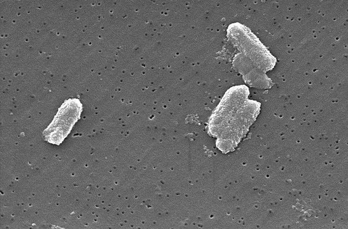
Most bacteria are nearly invisible under a regular light microscope because they lack color. To make them visible, microbiologists use stains—chemical dyes that color cells so their shape and structure can be studied.
Staining is simple, affordable, and fast. It helps scientists see bacterial size, shape, and arrangement clearly. Some stains even highlight special features like flagella, capsules, and spores.
What Are Stains?
In microbiology, stains (or dyes) are chemicals that contain chromophores, the parts that give color.
Most stains are salts—one part carries color while the other does not.
For instance, methylene blue chloride breaks apart in water into:
- A blue, positively charged methylene ion (the color part), and
- A colorless chloride ion.
The chemical structure of the dye determines which microorganisms it will react with.
Classification of Stains
1. Based on Method of Use
Microbiological stains are grouped into two main types:
- Simple stains: Use a single dye to color all microbes the same way.
- Differential stains: Use two or more dyes to highlight differences between types of bacteria or cell parts.
Simple stains such as methylene blue, safranin, carbol fuchsin, crystal violet, and India ink help visualize general features—size, shape, and arrangement.
Differential stains (like Gram, Acid-Fast, and Endospore stains) reveal deeper details, such as whether bacteria have thick or thin cell walls.
2. Based on Chemical Nature
- Basic stains: Have positively charged ions that bind to negatively charged bacterial components.
- Acidic stains: Contain negatively charged ions that color the background, not the bacteria.
- Neutral stains: Rarely used; India Ink is an example.
⚠️ Safety Note
Most biological dyes used in labs are aniline dyes derived from coal tar.
They can be carcinogenic, so always:
- Wear gloves and lab coats.
- Avoid skin contact and spills on benches.
- Handle dyes carefully and dispose of them properly.
General Staining Procedure
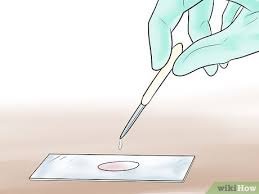
Before applying a stain, bacteria must be fixed to the slide so they don’t wash off.
Fixation Steps
- Prepare a thin smear of bacteria on a clean slide.
- Allow it to air-dry completely.
- Pass the slide (smear-side up) quickly through a flame 3–4 times.
The heat coagulates the proteins, helping bacteria stick to the slide.
However, overheating can distort cells, so apply just enough heat to fix them without burning.
Step-by-Step: Preparing a Bacterial Smear
- Clean the glass slide.
- Add a tiny drop of normal saline.
- Collect a small amount of bacterial culture and mix it with the drop until slightly cloudy.
- Spread the mixture thinly.
- Let it air-dry completely.
- Heat-fix gently by passing through a flame 3–4 times.
Common Stains and Their Applications
1. Romanowsky Stains
These include Wright’s, Leishman’s, Giemsa, and Jenner’s stains.
They combine eosin (a red dye) and methylene blue to color blood cells and parasites.
Used mainly in blood and bone marrow smears, these stains help identify malaria parasites and differentiate white blood cells.
2. Iodine
Used as a mordant in Gram staining, iodine helps the primary dye bind firmly to bacterial cell walls.
It also reacts with starch to produce a dark color, which is useful for some cell staining tests.
3. Crystal Violet
This purple dye binds strongly to bacterial cell walls and is the primary stain in the Gram method.
When combined with iodine, it forms a stable complex that turns Gram-positive bacteria deep purple.
4. Auramine and Rhodamine
These are fluorescent dyes used to detect acid-fast bacteria like Mycobacterium tuberculosis.
Under special light, Auramine-stained bacteria glow bright yellow or green, making detection easier.
Major Staining Techniques in Microbiology
- Methylene Blue Staining
- Gram Staining
- Albert Staining
- Ziehl-Neelsen (Acid-Fast) Staining
- India Ink Staining
- Iodine Staining for Ova and Cysts in Stool Samples
Quality Control of Stains
Always verify staining accuracy using control organisms.
For example:
| Stain | Control Organism | Expected Result |
|---|---|---|
| Gram | E. coli (–ve), S. aureus (+ve) | Purple cocci, pink rods |
| Ziehl-Neelsen | Mycobacterium spp., E. coli | Red bacilli, blue background |
| Iodine | Treated stool with cysts | Visible cyst nuclei |
Gram Staining: The Most Common Technique
Discovered by Hans Christian Gram in 1884, the Gram stain classifies bacteria into two groups:
- Gram-positive (thick cell wall, purple)
- Gram-negative (thin wall, pink)
Gram Stain Procedure
- Apply Crystal Violet for 1 minute, then rinse.
- Add Gram’s Iodine for 1 minute to form a dye complex.
- Wash briefly with ethanol or acetone—this removes the dye from Gram-negative cells.
- Counterstain with Safranin for 30 seconds to color Gram-negative bacteria pink.
Result:
- Gram-positive = Purple
- Gram-negative = Pink
Always include a positive and negative control to ensure accuracy.
Ziehl-Neelsen (Acid-Fast) Staining
This method identifies acid-fast bacilli (AFB) like Mycobacterium tuberculosis and M. leprae.
Reagents Used
- Carbol fuchsin (primary stain)
- 25% sulphuric acid (decolorizer)
- Methylene blue (counterstain)
Procedure
- Prepare and fix a smear from sputum.
- Flood with Carbol Fuchsin, heat gently for 5 minutes.
- Rinse and decolorize with acid for 2–4 minutes.
- Counterstain with Methylene Blue for 1 minute.
Result:
- Acid-fast bacilli → Red
- Non-acid-fast → Blue
India Ink Staining
Used mainly for detecting capsules in fungi such as Cryptococcus neoformans.
How It’s Done
- Place a drop of India ink on a slide.
- Mix a small amount of sample (e.g., cerebrospinal fluid) into the ink.
- Add a coverslip and view under the microscope.
The capsule appears clear against a dark background—an easy way to spot encapsulated organisms.
Conclusion
Staining is the foundation of medical microbiology.
By coloring invisible microbes, it helps scientists identify bacteria, diagnose infections, and guide treatment.
Always handle stains safely, follow proper procedures, and use controls to ensure reliable results.


Post Comment