Gram stain in the most interesting way this 2024!
“Come In and Stain!” – A Memorable Journey with Gram Staining 
1. The Basics: What Is Gram Staining?
Gram staining is like a microbial fashion show, where bacteria strut their stuff in vibrant colors. Developed by Danish physician Hans Christian Gram in 1884, this technique helps us categorize bacteria based on their cell wall composition. It’s like sorting them into two exclusive clubs: the Gram-Positives (the purple-loving crowd) and the Gram-Negatives (the red-and-pink enthusiasts).
2. The Players: Crystal Violet, Iodine, Alcohol, and Safranin
- Crystal Violet: Our primary stain, a dark blue-purple dye. It’s like the VIP pass to the Gram party.
- Iodine: The mordant. It forms a complex with crystal violet, making sure everyone stays in line.
- Alcohol (Decolorizer): The bouncer. It kicks out the unruly Gram-Negatives, leaving the Gram-Positives behind.
- Safranin: Our counterstain, a red dye. It’s like the after-party glow-up.
3. The Steps: How to Gram Stain Like a Pro

1; Gram stain procedure steps. from https://theory.labster.com/steps-gramstain/
- Preparation of the Smear:
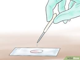
- Imagine you’re preparing a microscopic masterpiece. Clean your slide, draw two circles, and add a drop of water to each.
- Smear a tiny bit of bacteria (use aseptic technique!) within the circles. Air-dry and heat-fix the slide (but don’t cook the bacteria!).
- Staining Procedure:
- Crystal Violet: Flood the slide with crystal violet for 1 minute. Bacteria turn blue-purple.
- Iodine: Add Gram’s iodine for another minute. It’s like adding starch to thicken the sauce.
- Decolorization: Here’s the twist! Alcohol washes away the Gram-Negatives’ party vibes. They become colorless, while Gram-Positives hold onto their blue hue.
- Safranin: Finish strong with safranin. Gram-Negatives blush pink, but Gram-Positives stay true to their blue selves.
4. The Art of Interpretation
- Purple Lovers: Gram-Positives. Their thick peptidoglycan walls hold onto crystal violet.
- Red-and-Pink Enthusiasts: Gram-Negatives. Their thin walls let the dye escape during decolorization.
5. Exceptions to the Rule
- Intracellular Bacteria (e.g., Chlamydia): They’re the party crashers who hide inside host cells.
- Wall-Less Rebels (e.g., Mycoplasma): They skipped the cell wall class.
- Microscopic Celebrities (e.g., Spirochetes): Too tiny for light microscopy paparazzi.
6. Quality Control and Limitations
 Remember, Gram staining isn’t foolproof. Some bacteria defy the rules.
- But fear not! Armed with your newfound Gram knowledge, you’ll decipher bacterial secrets like a microbial Sherlock.
So, dear students, next time you encounter a slide, channel your inner Gram and decipher the colorful tales of bacteria. 🌟










Post Comment